Capillaroscopy
Observation of nailfold capillaries to support the assessment of autoimmune and vascular diseases.
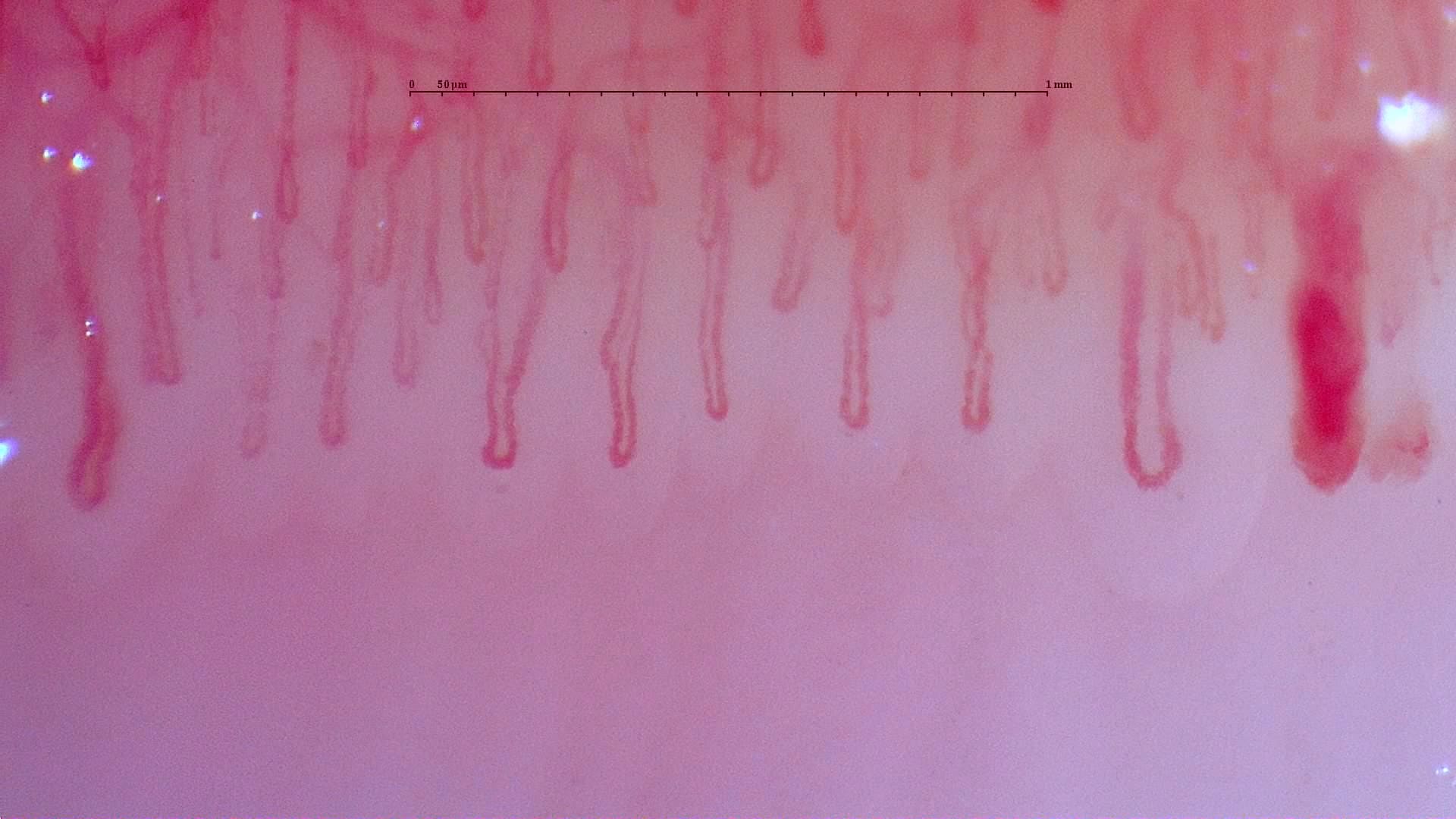
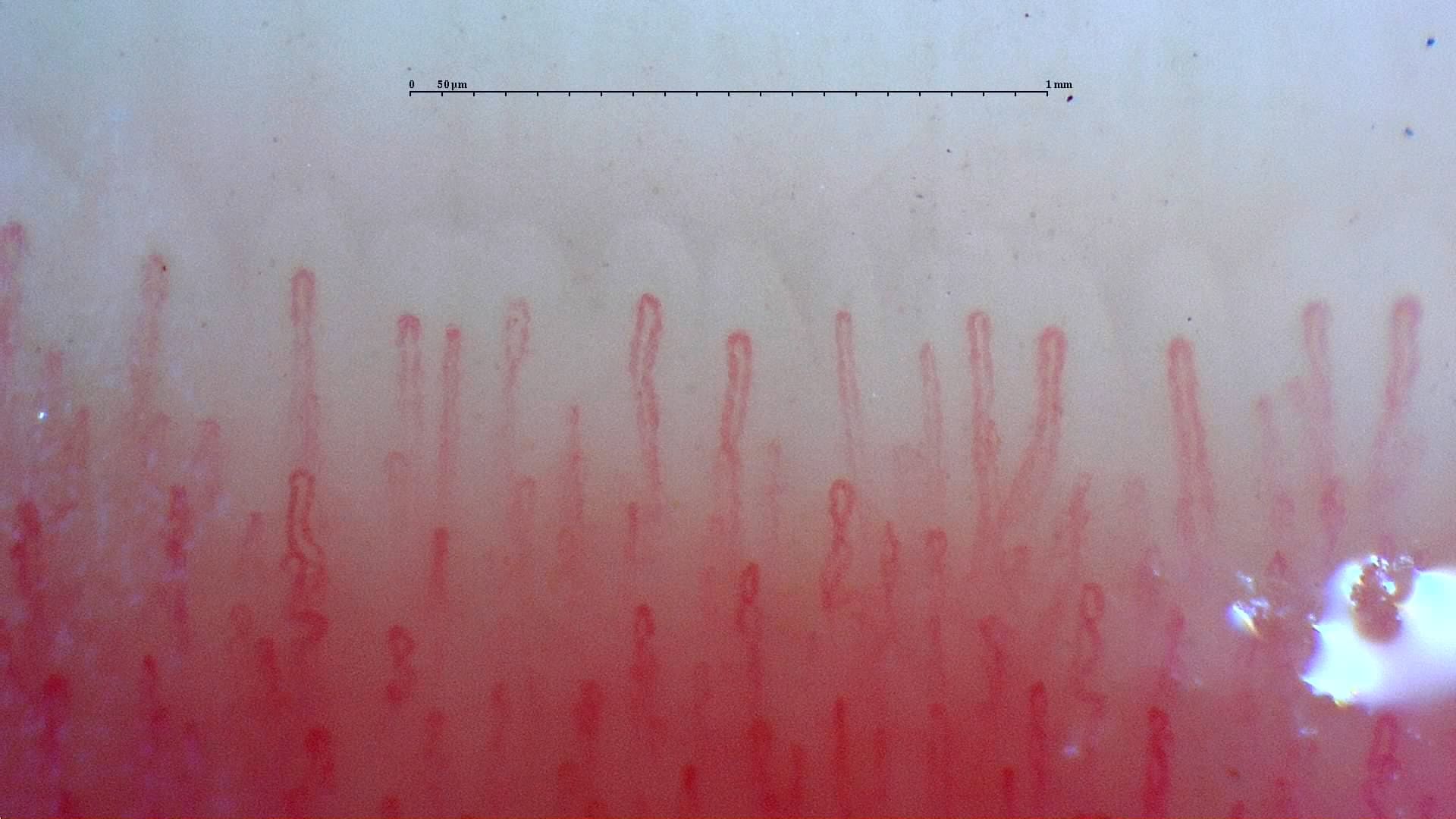
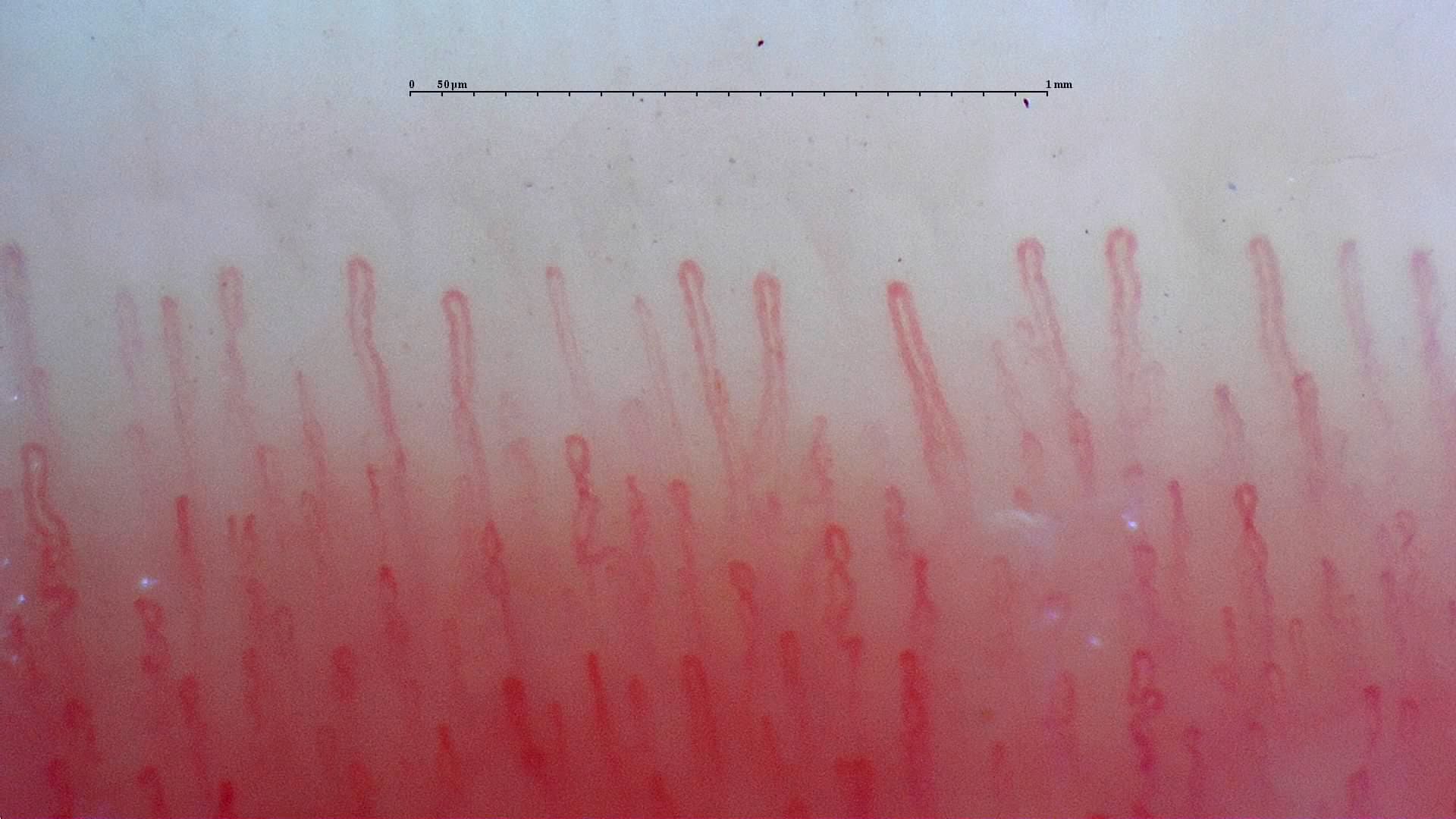
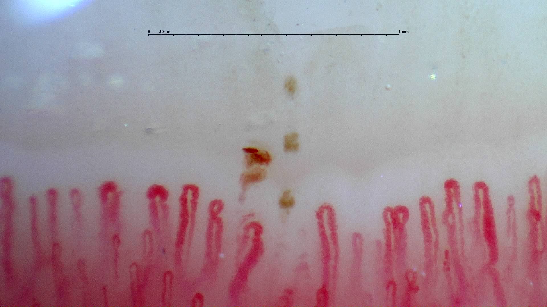
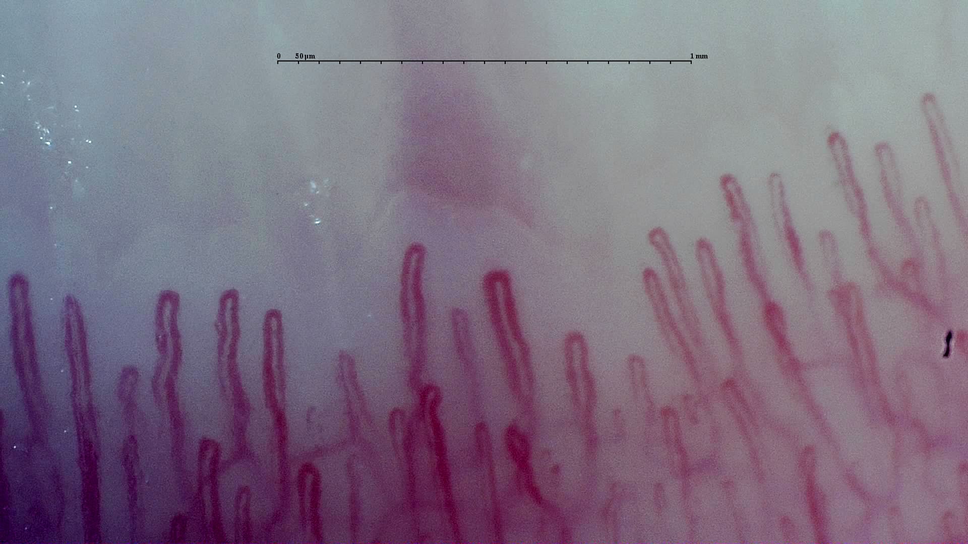
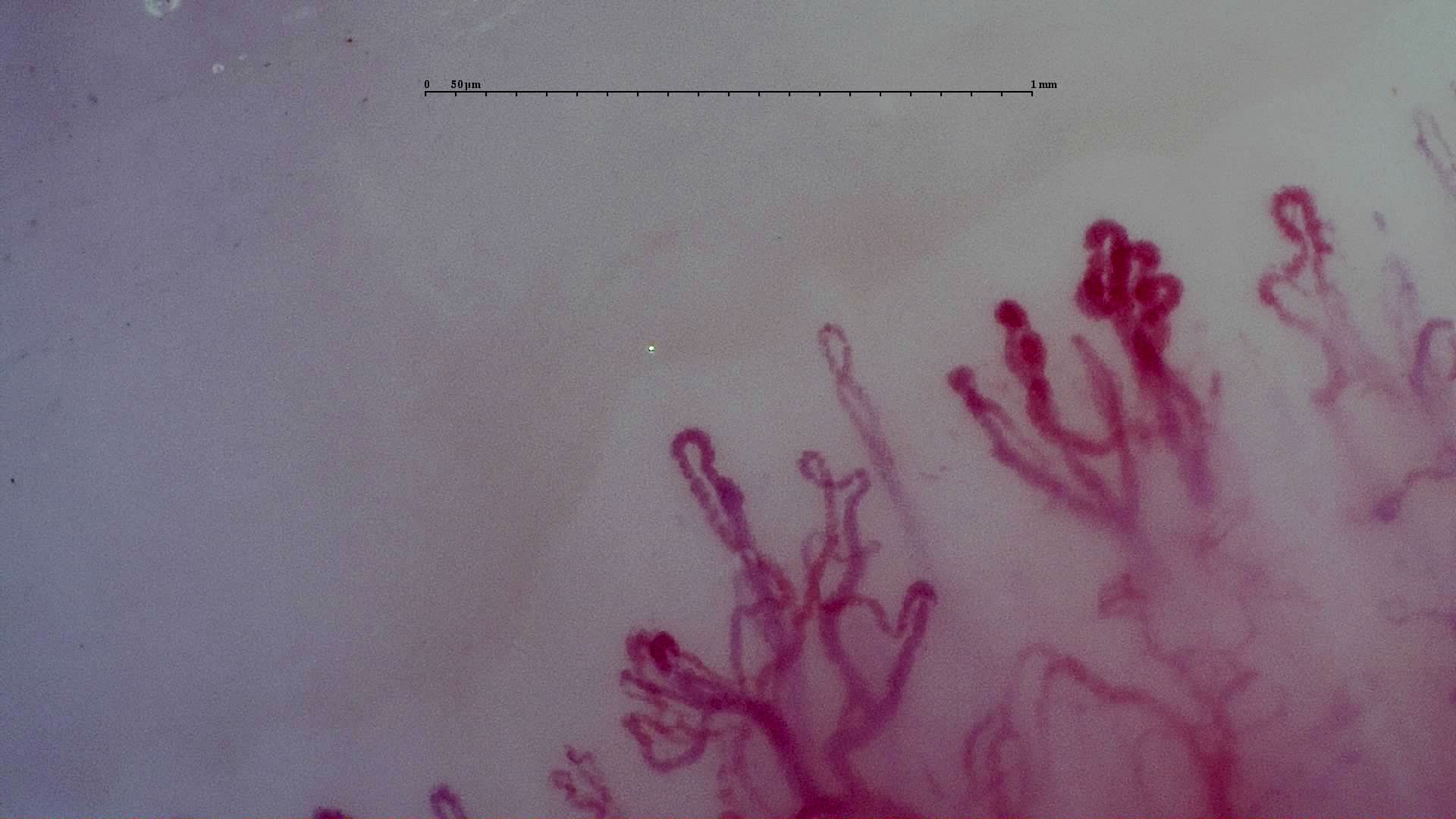
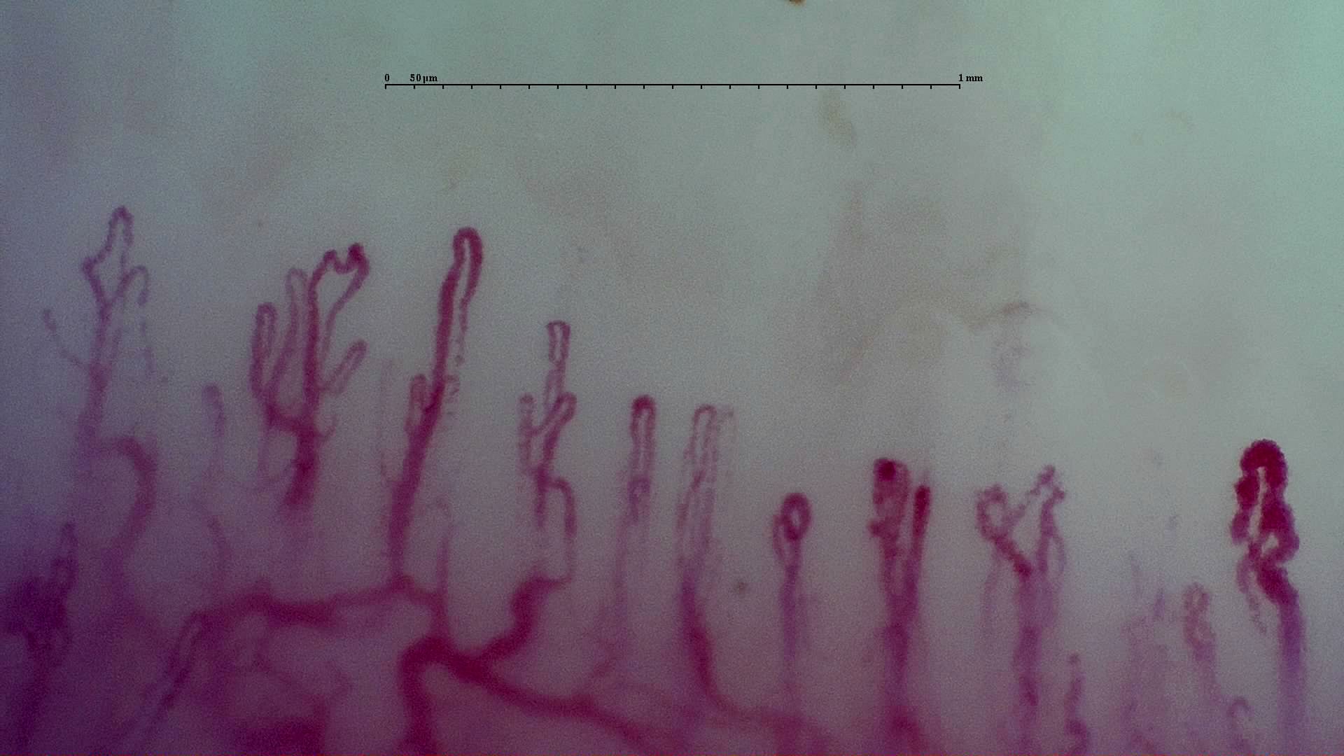
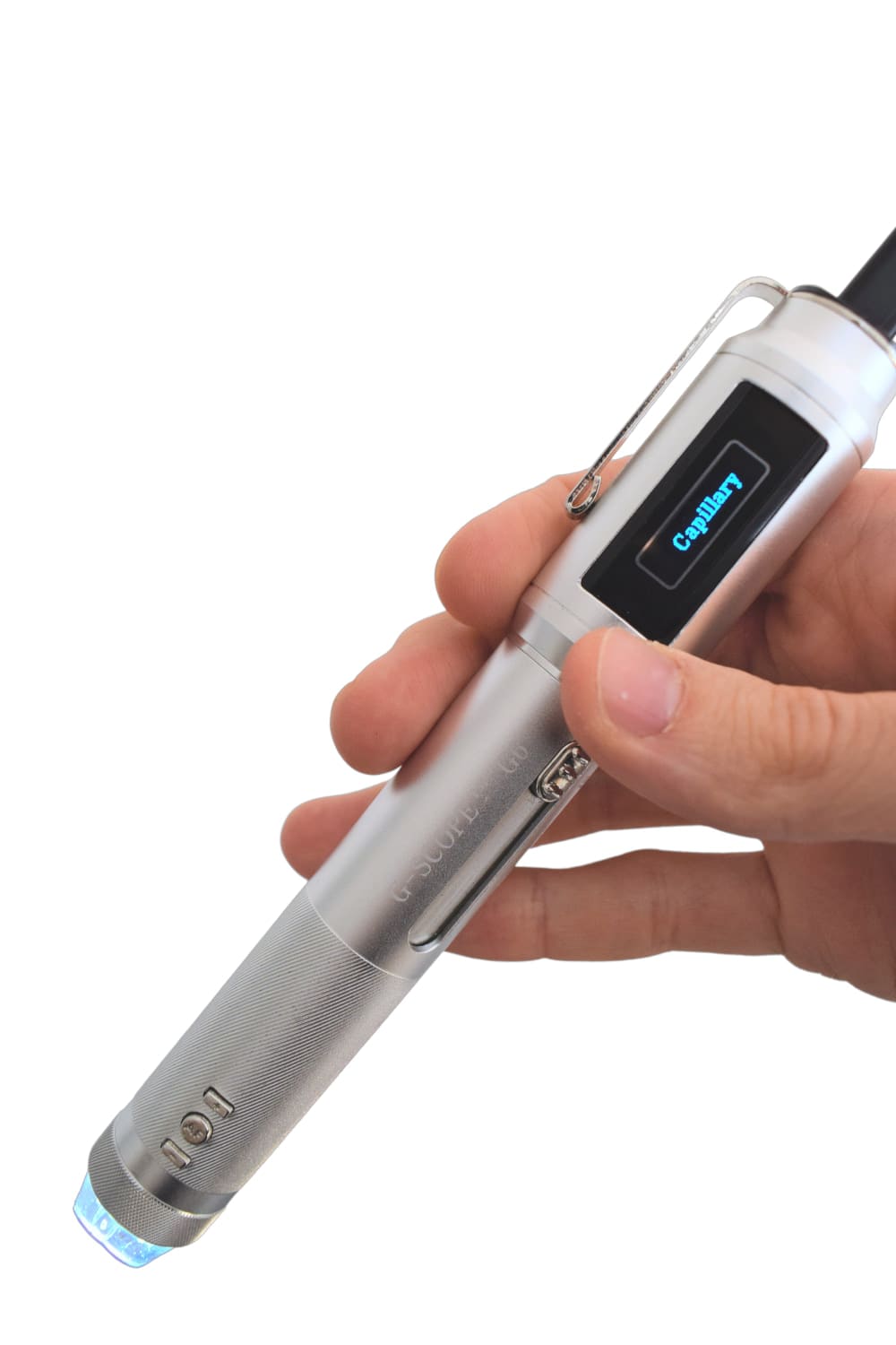
Nailfold capillaroscopy is an essential technique in rheumatology and internal medicine to assess microcirculation. When equipped with the dedicated capillaroscopy head (“capillary”), Smart G-Scope enables visualization of nailfold capillaries at 250× magnification, which is required for proper capillaroscopic assessment.
The capillaroscopy head is specifically designed for this application and is only functional at 250×. Its optical configuration allows detailed observation of capillary morphology when combined with proper contact technique.
For optimal visualization, an immersion medium must always be used in direct contact with the skin. Commonly used options include mineral oil or almond oil, which reduce surface reflections and improve capillary transparency.
Using this setup, clinicians can identify pathological patterns such as giant capillaries, microhemorrhages, avascular areas, and abnormal branching, supporting the assessment of systemic sclerosis, dermatomyositis, lupus, or primary versus secondary Raynaud’s phenomenon.
Documenting capillaroscopic findings with images and video supports longitudinal follow-up and interdisciplinary communication among rheumatologists, internists, and dermatologists.
Smart G-Scope Capillaroscope is fully compatible with Capillary.io CapillaryScope, a free desktop application specifically designed for nailfold capillaroscopy using USB capillaroscopes.
Key benefits
- Detailed visualization of nailfold capillary morphology at 250×
- Dedicated capillaroscopy head designed specifically for nailfold examination
- Improved image quality through mandatory immersion oil contact
- Support for recognizing scleroderma patterns and microangiopathy
- Photographic documentation for clinical records and follow-up
Image gallery
Click any image to enlarge
Practical examples
Early systemic sclerosis screening
A rheumatologist examines patients with Raynaud’s phenomenon using Smart G-Scope with the capillaroscopy head and immersion oil. The presence of giant capillaries and microhemorrhages supports early referral for immunologic work-up and systemic sclerosis confirmation.
Vasculitis monitoring
In patients with known vasculitis, periodic capillaroscopic follow-up using the capillary head at 250× allows clinicians to monitor treatment response by documenting changes in hemorrhages and capillary architecture.
Limitations & best practices
- ❗ Requires the dedicated “capillary” head, functional only at 250×. Make sure you purchase the capillaroscopy package.
- Immersion oil (e.g., mineral oil or almond oil) must always be used in contact with the skin.
- Does not replace serology or biopsy for definitive diagnosis.
- Stable positioning is required to avoid motion blur during capture.
Interested in this use case?
Buy Smart G-Scope
Get the right kit for this application. Secure checkout and fast shipping.
Need help choosing? Tell us your workflow and we’ll recommend the best setup.
Related use cases
Dermatology
High-precision visual analysis of skin lesions, nevi, and dermatologic changes.
Trichoscopy
Scalp and hair follicle examination to support the diagnosis of alopecia and hair disorders.
Aesthetics
Skin analysis, treatment evaluation, and documentation for professional aesthetic clinics.
Veterinary
Assessment of skin conditions, parasitoses, and hair analysis in companion and exotic animals.
Podiatry
Examination of nails, plantar skin, warts, and foot lesions for assessment and follow-up.